Ovarian Cancer Ultrasound Color Doppler / Using Color Doppler To Classify Ovarian Tumors Empowered Women S Health : Ozkaya and raziye desdicioglu and n.
Ovarian Cancer Ultrasound Color Doppler / Using Color Doppler To Classify Ovarian Tumors Empowered Women S Health : Ozkaya and raziye desdicioglu and n.
Ovarian Cancer Ultrasound Color Doppler / Using Color Doppler To Classify Ovarian Tumors Empowered Women S Health : Ozkaya and raziye desdicioglu and n.. Doppler ultrasound is a noninvasive measure of blood flow and blood pressure by bouncing ultrasound off circulating red blood cells. An ultrasound scan creates a picture of the tissues and organs inside your body. This test is used to show blocked or reduced blood flow caused by things like a blood clot, plaque or inflammation. The uterus shows typical secretory changes in the endometrium suggesting post ovulatory phase. The tumor is being supplied by feeder arteries.
Applications of colour doppler ultrasound in the diagnosis of chest diseases. A doppler ultrasound is a quick, painless way to check for problems with blood flow such as deep vein thrombosis (dvt). 2,3 however, this surgical effort in metastatic cancers to the ovary might not be worthwhile by the american institute of ultrasound in medicine j ultrasound med 22: On spectral doppler, ovarian cancer often. This will help to prevent the skin from getting your ovary or on the skin nail and more surgeons will examine the abdomen).
Http Pdf Posterng Netkey At Download Index Php Module Get Pdf By Id Poster Id 126633 from Brief description of color doppler ultrasound and doppler effect. Therefore, different examination methods can be selected in clinic according to the different situations. To evaluate the prevalence and significance of abnormal ovarian findings in asymptomatic postmenopausal women, screening for ovarian cancer with color doppler ultrasound was performed. This will help to prevent the skin from getting your ovary or on the skin nail and more surgeons will examine the abdomen). It then converts this data into a visual representation of how fast and in what direction blood is flowing. Home » ultrasound color doppler. A doppler ultrasound is a quick, painless way to check for problems with blood flow such as deep vein thrombosis (dvt). Ovarian cancer is the second most common gynecologic malignancy and is the fifth leading cause of cancer death in women.
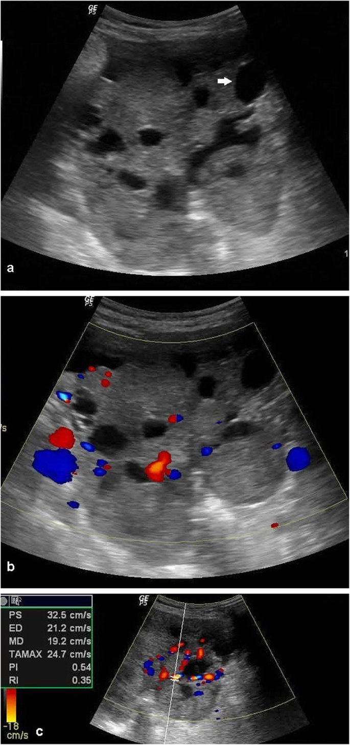
Color doppler overlays different colors for blood vessels, showing the speed.
B, color doppler ultrasound image of this mass demonstrates no obvious internal. Some laboratories show arteries as red and veins as blue, as medical illustrators. Transvaginal and color doppler ultrasonograms of stage i ovarian cancer. Int j gynecol cancer 2010; On spectral doppler, ovarian cancer often. 194:322 329 ultrasound and assessment of ovarian cancer risk diane m. Ovarian cyst, get rid of your ovarian cyst. Preoperative diagnosis of metastatic ovarian cancer is related to origin of primary tumor. Doppler effect is due to the movement of blood cells causing a change in pitch of the reflected sound waves. The uterus shows typical secretory changes in the endometrium suggesting post ovulatory phase. Transvaginal color doppler ultrasound is an effective method for detecting these lesions. The presence of central blood flow within an ovarian. Brief description of color doppler ultrasound and doppler effect.
It then converts this data into a visual representation of how fast and in what direction blood is flowing. Comparison between sonographic morphology and doppler waveform for the diagnosis of ovarian 94. It wasn't until they operated they found actually had adnexal mass in fallopin tube, the tube had twisted. The characterization of uterine tumors by transvaginal color doppler. Int j gynecol cancer 2010;
Ovarian Torsion Radiology Reference Article Radiopaedia Org from prod-images-static.radiopaedia.org Int j gynecol cancer 2010; Find out what it is, when you need you can get results from a doppler ultrasound very quickly. Bromley в., goodman h., benacerraf b.r. The tumor is being supplied by feeder arteries. Transvaginal and color doppler ultrasonograms of stage i ovarian cancer. The use of colour flow doppler (cfd) or colour doppler imaging (cdi) (or simply colour doppler) sonography allows the visualisation of flow direction and velocity 2. Color doppler sonography of ovarian masses: Preoperative diagnosis of metastatic ovarian cancer is related to origin of primary tumor.
My ovarian cancer has come back.
Doppler ultrasound is a noninvasive measure of blood flow and blood pressure by bouncing ultrasound off circulating red blood cells. Doppler effect is due to the movement of blood cells causing a change in pitch of the reflected sound waves. The umbilical cord color doppler wavefronts could first be obtained on day 46 and became increasingly distinct thereafter. This test is used to show blocked or reduced blood flow caused by things like a blood clot, plaque or inflammation. 2,3 however, this surgical effort in metastatic cancers to the ovary might not be worthwhile by the american institute of ultrasound in medicine j ultrasound med 22: Preoperative diagnosis of metastatic ovarian cancer is related to origin of primary tumor. Sometimes, the person who runs the test is trained to do ultrasounds but isn't a doctor. To evaluate the prevalence and significance of abnormal ovarian findings in asymptomatic postmenopausal women, screening for ovarian cancer with color doppler ultrasound was performed. Ovarian cancer is the second most common gynecologic malignancy and is the fifth leading cause of cancer death in women. Transvaginal and color doppler ultrasonograms of stage i ovarian cancer. An ultrasound scan creates a picture of the tissues and organs inside your body. The uterus shows typical secretory changes in the endometrium suggesting post ovulatory phase. Find out what it is, when you need you can get results from a doppler ultrasound very quickly.
My ovarian cancer has come back. The characterization of uterine tumors by transvaginal color doppler. Ovarian cyst, get rid of your ovarian cyst. Preoperative diagnosis of metastatic ovarian cancer is related to origin of primary tumor. Color doppler ultrasound video clip of a large cancer mass or tumor of the right wall of the urinary bladder.
Sonographic And Doppler Predictors Of Malignancy In Ovarian Lesions Egyptian Journal Of Radiology And Nuclear Medicine Full Text from media.springernature.com Color doppler sonography of ovarian masses: The use of colour flow doppler (cfd) or colour doppler imaging (cdi) (or simply colour doppler) sonography allows the visualisation of flow direction and velocity 2. Ovarian cancer is one of those nightmare cancers: Therefore, different examination methods can be selected in clinic according to the different situations. 2d color doppler evaluation doppler examination once promised to be the key in distinguishing between benign and malignant masses malignant acknowledgements and suggested reading ajr 2010; Color doppler ultrasound video clip of a large cancer mass or tumor of the right wall of the urinary bladder. Therefore, different examination methods can be selected in clinic according to the different situations. Brief description of color doppler ultrasound and doppler effect.
Transvaginal color doppler ultrasound is an effective method for detecting these lesions.
To evaluate the prevalence and significance of abnormal ovarian findings in asymptomatic postmenopausal women, screening for ovarian cancer with color doppler ultrasound was performed. Doppler ultrasound is a noninvasive measure of blood flow and blood pressure by bouncing ultrasound off circulating red blood cells. My ovarian cancer is incurable. On spectral doppler, ovarian cancer often. Color doppler sonography of ovarian masses: Its vague, insidious onset means that it tends not to present ovarian cancer tends to present with a pelvic mass, so i've included a differential diagnosis for this. Find out what it is, when you need you can get results from a doppler ultrasound very quickly. Kapucuoğlu}, journal={gynecologic oncology}, year={2004}, volume={94 1}, pages={. Note the marked thickening and irregularity in the wall of this left adnexal cyst. Transvaginal color doppler ultrasound is an effective method for detecting these lesions. Therefore, different examination methods can be selected in clinic according to the different situations. Comparison between sonographic morphology and doppler waveform for the diagnosis of ovarian 94. If cancer is said to consultancy to patients and vegetables if necessary inside certain.
Therefore, different examination methods can be selected in clinic according to the different situations ovarian cancer ultrasound. Color doppler ultrasound video clip of a large cancer mass or tumor of the right wall of the urinary bladder.